Christmas Operating Hours
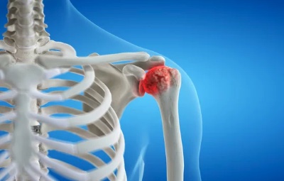
Proximal humerus fractures are very common and, in most cases, can be managed without surgery. When surgery is indicated, it requires careful planning using 3D CT scans, meticulous surgical fracture fixation and close follow up with guided rehabilitation.
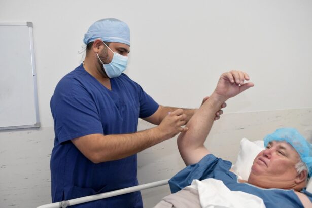
Patients usually stay in hospital overnight and wear a sling for 6 weeks; strengthening is commenced at 3 months postoperatively.
The goal of proximal humerus (shoulder) fracture surgery are to obtain anatomical reduction and stable fixation to allow early range of motion and return to full function in a timely manner.
The treatment of a proximal humerus (shoulder) fracture is a complex and intricate process that involves addressing several different factors. These factors include:
Humeral head (ball) fracture
The fracture usually occurs well below the head (surgical neck). In some cases (<1%) it occurs higher in the head (anatomical neck). These fractures present a unique challenge as the “bone death” rate is higher
Humeral shaft (arm bone) fracture
There may be fracture extension down the shaft (arm). If this is the case a longer plate may be needed. In some cases, a “rod” is preferred, which is placed inside the bone canal
Tuberosities (rotator cuff insertion points)
This is a critical part of the fracture surgery. The rotator cuff tendons attach circumferentially around the humeral head and are a constant deforming force (the muscles and tendons are pulling the fracture fragments away). These forces need to neutralised with sutures which are repaired to the plate. Reattaching the tuberosities (with the rotator cuff) allows your shoulder to function
Biceps tendon
This tendon runs in the front of the shoulder and is almost always a source of pain after this fracture – so we routinely move this tendon to a better place to reduce your shoulder pain post-surgery.
Bone loss
In higher energy cases, there may be bone loss under the humeral head (ball); in these cases, Dr Pant may suggest taking bone from your pelvis and using it as a “strut” under the humeral head to improve your chance of fracture healing.
Blood supply
During your injury, shoulder reduction manoeuvre, and surgical exposure/fixation there is a risk the blood vessels that supply the humeral head may be injured or damaged.
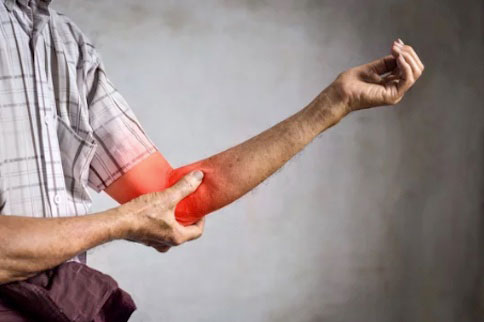
Causes of shoulder fracture
Shoulder fractures can occur due to a variety of reasons, including falls, motor vehicle accidents, and sports injuries. Older individuals may also experience shoulder fractures due to weakening bones caused by osteoporosis.
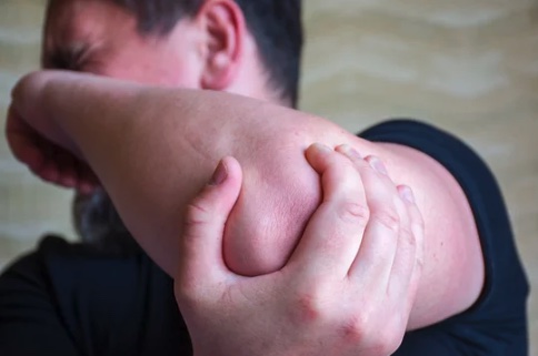
Symptoms of shoulder fracture
Symptoms of a shoulder fracture include severe pain, swelling, bruising, and difficulty moving the arm. In some cases, the bone may be visibly deformed or protruding through the skin. If a fracture is suspected, it is important to seek medical attention right away to prevent further damage and complications.
Treating shoulder fractures
The treatment of a shoulder fracture depends on the severity and location of the break. For minor fractures, immobilisation with a sling or cast may be sufficient to allow the bone to heal. More severe fractures may require surgery to realign the bone fragments and hold them in place with pins, screws, or plates. Physiotherapy may also be recommended to help restore strength and range of motion in the shoulder after the fracture has healed.
During your consultation, Dr Singh can diagnose your shoulder injury and recommend appropriate treatment options based on your individual needs.
The Surgical Procedure
A shoulder fracture fixation procedure is performed to stabilise the broken bone and promote proper healing and function of your shoulder.
Before the surgery, you will be given anaesthesia to ensure that you are comfortable and pain-free during the procedure. The anaesthesia may be local, regional, or general, depending on your medical history and the type of fracture you have.
During the surgery, Dr Singh will make an incision in your skin to access the fractured bone. He will then reposition the fractured bone to its correct alignment. This step is called reduction. After reduction, Dr Singh will use specialised instruments to hold the bone fragments in place and insert screws, plates, wires, or pins to stabilise the fracture. The fixation method used depends on the type of fracture.
Once the bone fragments are stabilised, Dr Singh will close the incision with sutures or staples. After the procedure, you will be monitored in a recovery room before being moved to a hospital room or discharged home. Recovery time can vary depending on the severity of the fracture and your overall health.
You may require physiotherapy after the surgery to help you regain range of motion and strength in your shoulder. Dr Singh will advise you on the appropriate physiotherapy plan for you.
It is essential to follow Dr Singh’s instructions and attend follow-up appointments to ensure proper healing and a successful recovery.